In 1895, Wilhelm C. Roentgen discovered X-rays and photographed the world's first X-piece, the photo of the palm of the ring. In 1967, Godfrey N. Hounsfield invented the first CT device, which was able to take pictures from multiple angles, capture the three-dimensional information of the object, and observe its internal structure without destroying the object. In the 1970s, hospitals began using CT to diagnose diseases. For decades, this great technology has been widely used in various fields, such as medicine (tissue of tissues and organs, physiological metabolic processes), pharmacy (pharmaceutical testing, new drug development), materials science (development of new materials), and industry (each Quality inspection and flaw detection), agriculture (quality inspection and analysis of wood and seeds), engineering (internal porosity, connectivity and permeability analysis of building materials), jewelry (authentic identification and optimal cutting design), Archaeology (structure and composition analysis of fossils) and other fields.
The most well-known CT is the medical CT used for clinical examination. The first CT picture shows the skull image. After more than 40 years of development, Hounsfield's extremely slow translating pencil beam CT has evolved into a wide variety of CT families, such as spiral CT, 64-row volumetric CT, and quantitative CT.
Basic classification of CT equipment
Types of | FOV | Resolution | description |
CT | 10-60cm | 500-1500μm | Clinical CT is mainly based on human body scan, and the quantitative analysis software is installed to become QCT (quantitative CT). Since the invention of spiral CT, the scanning speed has been accelerated, and the whole body scan can be completed in a few minutes. However, it is difficult to increase the resolution due to the size of the FOV and the radiation dose. |
pQCT | 5-15cm | 50-500μm | Peripheral quantitative CT (peripheral Quantitative CT) scans the limbs of the human body and can be used for clinical diagnosis and scientific research. pQCT can analyze trabecular bone and cortical bone separately, and can perform biomechanical analysis to accurately predict fracture risk. Without the influence of receptor position, body shape and bone hyperplasia, the risk assessment of osteoporosis has obvious advantages over DEXA. |
microCT | 1-8cm | 5-80μm | Micro-CT, using micro-focus X-ray tube, high resolution, but small imaging range for scientific research. Including in vitro ( in vitro ) and in vivo ( in vivo ), the former for bone and other specimens, the latter for living small animal scanning. |
CTM | 0.01-0.5cm | 0.1-10μm | X-Ray Computerized Tomography Microscopy, parallel X-ray imaging produced by a synchrotron. The highest resolution, sub-micron, but the FOV is extremely small. Single-energy X-ray, high image quality. |
In the 1980s, due to the inability of ordinary CT to meet the demanding requirements of scientific research, the academic community began to develop micro-CT, MicroCT. MicroCT (also known as micro-CT, micro-focus CT or micro-CT) uses a micro-focus X-ray tube that is different from ordinary clinical CT with a resolution of up to a few microns, second only to the level of synchronous accelerated X-ray imaging equipment. Has a good "microscopic" effect. The price paid for the high resolution is that the volume of the scanned sample is very small, only a few centimeters, reflecting its "mini" side.
Unlike the Fan Beam, which is commonly used in clinical CT, the MicroCT usually uses a Cone Beam. The use of a cone beam not only enables true isotropic volumetric images, improves spatial resolution, improves ray utilization, but is much faster than fan beam CT when acquiring the same 3D image.

MicroCT imaging principle
Two basic types of information that MicroCT can provide: geometric information and structural information. The former includes the size, volume, and spatial coordinates of each point, and the latter includes material information such as the attenuation value, density, and porosity of the sample. In addition, SCANCO's finite element analysis function can also provide mechanical parameters such as elastic modulus and Poisson's ratio of the tested materials, analyze the stress and strain of the sample, and perform non-destructive mechanical tests.
The MicroCT 2 Basic structure
l Sample stationary, X-ray tube and detector movement: This structure is consistent with the clinical spiral CT, and the bulb rotates around the sample. The scanning speed is fast, the radiation dose is small, and the spatial resolution is low, which is mostly used for living animal scanning.
l Sample movement, X-ray tube and detector fixation: The sample spins between the tube and the detector, and can be moved up and down and back and forth. The scanning speed is slow, the radiation dose is large, and the spatial resolution is high, which is mostly used for scanning excised specimens.
The MicroCT 2 Class application object
l In vivo : The study subjects are usually small animals such as mice, rats or rabbits, which are anesthetized or fixed and scanned. Longitudinal studies of physiological metabolic functions can be achieved, significantly reducing the number of animals required for animal testing. Similar to medical clinical CT, the living small animal MicroCT is also capable of respiratory gating and enhanced scanning (using contrast agents).
l in vitro : The study object is usually an ex vivo specimen (such as bones, teeth) or samples of various materials, analysis of internal structure and mechanical properties. It is also possible to perfuse live animals with a coagulated contrast agent for fine imaging of the cardiovascular, urinary or digestive systems.
Main application areas of MicroCT
l bones. Bone is one of the most important applications of MicroCT, and trabecular bone is the main research object. Changes in cancellous and cortical bone are associated with conditions such as osteoporosis, fractures, osteoarthritis, ischemia, and genetic diseases. At present, MicroCT technology has largely replaced the destructive histomorphometry method.
l bones. Bone is one of the most important applications of MicroCT, and trabecular bone is the main research object. Changes in cancellous and cortical bone are associated with conditions such as osteoporosis, fractures, osteoarthritis, ischemia, and genetic diseases. At present, MicroCT technology has largely replaced the destructive histomorphometry method.
l teeth and periodontal tissue. From the 3D overall structure, it is possible to study the changes of root canal morphology, caries destruction, dental tissue density changes, alveolar bone structure and mechanical properties.
l Biological materials. For example, the parameters such as porosity and strength of the biomimetic material scaffold prepared in vitro are analyzed, the scaffold design is optimized, the tissue sample to be replaced is scanned, the three-dimensional image is acquired, and the output is an STL file for rapid prototyping (CAD/CAM), and the like.
l disease mechanism research. For example, study the effects of different genes or signaling pathways on the quantity or quality of bones, the effects of disease states on bone development/repair, evaluate the effects of hyperlipidemia on valvular calcification, and the cytokine growth of blood vessels after repair of tissue after fracture. Influence, and so on.
l New drug development. For example, to study new osteoporosis drugs and efficacy evaluation, MicroCT has been called an important preclinical detection technique.
l Other. Quality inspection and flaw detection of micro-devices, internal porosity, connectivity and permeability analysis of building materials, authenticity identification of jewelry and optimal cutting scheme design, and analysis of fossil structure.
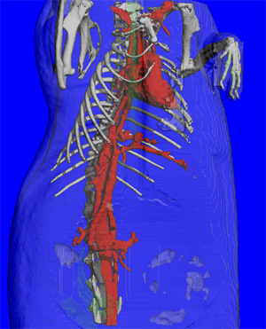 |
Thoracic imaging of living mice (volume reproduction) |
Sodium Gluconate,High Quality Sodium Gluconate,Sodium Gluconate Details
SHANDONG BAISHENG BIOTECHNOLOGY COM , https://www.baishengbioproducts.com